
Why You Stop Sleeping Deeply After 40 — And What the Growth Hormone Pulse Has to Do With It
Your deep sleep and your growth hormone don't decline separately — they decline together. Here's the mechanism most physicians never explain, and what the data shows about restoring it.
You go to bed. You wake up six hours later. You were technically asleep the whole night — but you don't feel rested. Your joints are stiff. Your mind is slow. Recovery from your workouts has gotten noticeably worse. And nobody has a good explanation.
Here's what most physicians don't connect: deep, restorative sleep and growth hormone secretion are not two separate things. They are the same system — and that system degrades quietly, predictably, starting in your late thirties.
Understanding this mechanism is the first step toward doing something about it.
The Architecture of Sleep — and Where Growth Hormone Lives
Sleep is not a single state. It moves through cycles — typically four to six per night — alternating between lighter stages and a specific phase called slow-wave sleep (SWS), also referred to as Stage 3 or deep NREM sleep. This is the stage characterized by long, rolling delta waves on an EEG. It is the most physically restorative part of the sleep cycle.
Here is what makes slow-wave sleep biologically distinct: it is precisely synchronized with the largest growth hormone pulse of the day.
Within the first 90 minutes after falling asleep, the hypothalamus releases a surge of growth hormone-releasing hormone (GHRH). GHRH activates both the pituitary — triggering GH secretion — and specific sleep-promoting neurons in the preoptic area of the hypothalamus, including the ventrolateral preoptic nucleus (VLPO). These neurons drive slow-wave sleep directly. The result: a single cascade generates both the GH pulse and the deep sleep stage simultaneously.
This is not coincidental overlap. It is bidirectional coupling. More slow-wave sleep produces a larger GH pulse; a larger GH pulse deepens subsequent slow-wave sleep. The two reinforce each other throughout the early part of the night.
That first nocturnal GH pulse often accounts for 50 to 70 percent of the body's total daily growth hormone output. It is not a minor event — it is the primary anabolic signal of the 24-hour cycle.
What Happens to This System as You Age
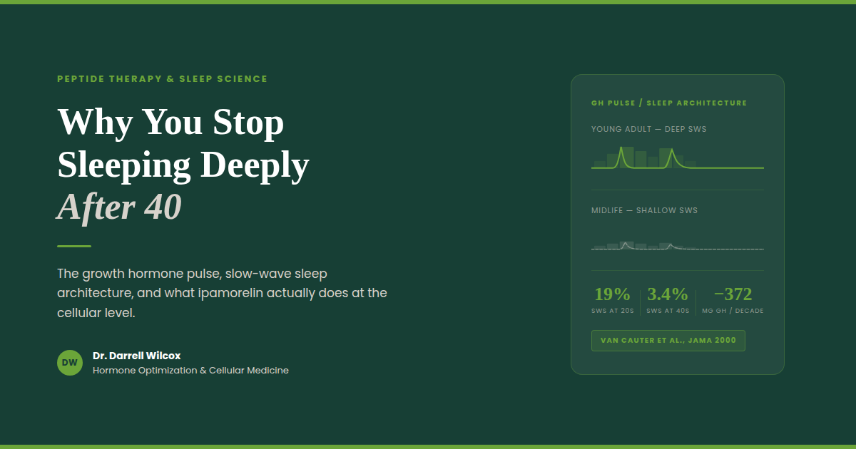
A landmark study published in JAMA tracked sleep architecture and GH secretion across the lifespan in healthy men and found a pattern that is difficult to ignore.
Deep slow-wave sleep accounted for roughly 19 percent of total sleep time in early adulthood. By midlife — ages 36 to 50 — it had dropped to approximately 3.4 percent. Growth hormone secretion tracked that decline almost exactly, falling by more than 370 micrograms per decade from early adulthood through midlife. The association between slow-wave sleep and GH output was statistically significant independent of age: less deep sleep meant less growth hormone, regardless of how old the person was.
This is the somatopause — the gradual age-related decline of the GH axis — playing out at the level of your sleep architecture.
The consequences are clinical. Less GH during sleep means reduced protein synthesis overnight, slower tissue repair, impaired fat metabolism, and diminished recovery from exercise. It also means lighter sleep: the bidirectional loop works in both directions. When GH output drops, slow-wave sleep quality deteriorates further. The system doesn't just decline — it entrains toward a lower equilibrium.
By the time most people notice their sleep has gotten worse and their recovery has slowed down, the mechanism has been shifting for years.
Growth Hormone During Sleep — What It Actually Does
Understanding why sleep-associated GH matters requires a brief look at what growth hormone does downstream.
GH does not act directly on muscle and tissue in most cases. It signals the liver to produce insulin-like growth factor 1 (IGF-1), and it is primarily IGF-1 that drives the anabolic effects most people associate with growth hormone: muscle protein synthesis via the PI3K/Akt/mTOR signaling pathway, satellite cell recruitment for muscle repair, and suppression of protein degradation pathways. GH also acts directly on adipose tissue to promote lipolysis — particularly in visceral fat, which has a high density of GH receptors.
Sleep deprivation disrupts both sides of this equation. Research shows that insufficient or fragmented sleep elevates cortisol while suppressing GH and IGF-1. Cortisol activates muscle protein degradation pathways directly. The net effect is a shift toward a catabolic hormonal environment during the overnight hours — the opposite of what the body is designed to do during sleep.
For someone who trains consistently, the math is straightforward: if the anabolic signaling that drives overnight recovery is compromised, training adaptations slow. Not because of how hard you are working, but because of what is happening — or not happening — during the hours you are not working at all.
Ipamorelin: Restoring the Pulse Without Disrupting the Signal
Ipamorelin is a synthetic pentapeptide that acts as a selective agonist at the ghrelin receptor (GHS-R1a) on the anterior pituitary. It stimulates the pituitary to release growth hormone in a pulsatile pattern — the same physiologic pattern as endogenous GH secretion.
What makes ipamorelin clinically distinct is what it does not do.
Earlier growth hormone-releasing peptides — GHRP-2 and GHRP-6 — also stimulate GH through the ghrelin receptor, but they simultaneously activate non-GHS-R1a pathways that drive elevations in ACTH and cortisol. In a 1998 study that established ipamorelin as a separate category, researchers found that GHRP-2 and GHRP-6 produced significant increases in cortisol and ACTH alongside their GH-releasing effects. Ipamorelin, administered at comparable doses, produced no statistically significant elevation in ACTH or cortisol — even at doses more than 200 times the amount required for maximal GH release. It also had no effect on prolactin, FSH, LH, or TSH.
This selectivity matters clinically. Cortisol is catabolic. A GH secretagogue that raises cortisol alongside GH is working against the recovery environment you are trying to create. Ipamorelin amplifies GH output through the ghrelin receptor while leaving the rest of the hormonal landscape undisturbed.
Administered in the evening, ipamorelin is timed to coincide with the natural nocturnal GH pulse — reinforcing the signal rather than overriding it. The result is an amplified slow-wave sleep-coupled GH release, which supports deeper sleep architecture and enhances the downstream anabolic and tissue repair processes that follow.
Ipamorelin does not introduce synthetic growth hormone. It signals your pituitary to produce more of its own — in the same pulsatile pattern your body was designed to use.
Other Peptides That Work on the Same Axis — and How They Differ
Ipamorelin is one tool in a larger pharmacological toolkit. Other peptides operate on the same GH axis through different receptor pathways, with different clinical profiles.
Sermorelin is a 29-amino-acid analog of GHRH — the upstream signal that the hypothalamus sends to the pituitary to initiate GH release. It is often administered at bedtime to align with the natural nocturnal pulse. Because it works at the GHRH receptor rather than the ghrelin receptor, its GH-releasing effect is subject to somatostatin regulation: when somatostatin levels are high, sermorelin's effect is blunted. It has a relatively short half-life and produces a more transient GH pulse than longer-acting analogs.
Ipamorelin combined with a GHRH analog (such as modified GRF 1-29, sometimes referred to as CJC-1295 without DAC) represents a complementary approach. The GHRH analog activates the GHRH receptor — amplifying the amplitude of each GH pulse. Ipamorelin activates the ghrelin receptor — triggering pulse initiation with high selectivity. These are distinct receptor pathways converging on the same output. A 2006 randomized, placebo-controlled trial confirmed that subcutaneous CJC-1295 produced sustained, dose-dependent increases in GH and IGF-1 in healthy adults, with an estimated half-life of 5.8 to 8.1 days and no serious adverse reactions reported. A companion analysis by Ionescu and Frohman, also in 2006, confirmed that pulsatile GH secretion was preserved throughout continuous GHRH stimulation — GH continued to be released in discrete pulses rather than continuously.
Tesamorelin is a 44-amino-acid GHRH analog with an added structural modification that increases its stability and half-life compared to endogenous GHRH. It is the only peptide in this class that is FDA-approved — specifically for reducing excess visceral abdominal fat in patients with HIV-associated lipodystrophy. Its mechanism is the same GHRH receptor pathway as sermorelin, but with greater potency and duration. In non-HIV populations, it is used off-label for metabolic optimization and body composition. Like sermorelin and CJC-1295, its GH-stimulating effect occurs upstream — it tells the pituitary to release GH rather than supplying GH directly.
The choice among these peptides — and whether they are combined — depends on the individual's GH axis status, metabolic goals, and clinical presentation. No single protocol fits every patient.
What to Expect — Realistic Timeline and Monitoring
Patients who begin ipamorelin-based protocols typically report improvement in sleep quality within two to four weeks — most commonly describing it as feeling more rested after the same hours of sleep, or waking less frequently during the night. Body composition changes, if present, tend to emerge over a longer window of eight to twelve weeks, as improved overnight GH signaling supports changes in fat metabolism and lean tissue preservation.
IGF-1 is the standard monitoring marker. Because these peptides work by amplifying endogenous GH production, IGF-1 rises proportionally. Baseline measurement before initiating therapy, followed by follow-up testing at six to eight weeks, allows for appropriate dose titration and confirms that the GH axis is responding as expected. If IGF-1 rises above age-appropriate reference ranges, dosing should be adjusted.
These peptides are not appropriate for individuals with active malignancy, given the theoretical concern about IGF-1's role in cell proliferation. This is an absolute contraindication. Individual response also varies based on age, baseline GH axis function, sleep quality at the time of initiation, and concurrent hormonal status.
A Note on Regulatory Status
Ipamorelin, sermorelin, and compounded CJC-1295 are not FDA-approved for the indications described here. Their use in adult patients for sleep, recovery, and body composition optimization is off-label. Tesamorelin (Egrifta) carries FDA approval specifically for HIV-associated lipodystrophy; off-label use in other populations requires clinical judgment and informed patient consent.
Compounded versions of these peptides are available through licensed compounding pharmacies and are prescribed by physicians operating within applicable state and federal regulations. Monitoring requirements apply to all protocols.
The Conversation Worth Having
If your sleep has quietly gotten worse over the last several years — if you wake up less rested, recover more slowly, and feel like the work you put in isn't translating the way it used to — the GH axis is worth evaluating.
Conventional medicine rarely looks at this. Sleep is treated as a lifestyle issue. Recovery is treated as a training issue. The hormonal architecture that connects the two is rarely measured and almost never optimized.
That is exactly where this conversation starts.
If you are interested in exploring whether peptide therapy is appropriate for your clinical picture, a virtual consultation is a good first step. We review your history, your symptoms, and where indicated, the relevant labs — and build a protocol around what your physiology actually needs.
Click the link in our bio to connect with us and schedule your free consultation.
[DISCLAIMER]
This post is for educational purposes only and does not constitute medical advice, diagnosis, or treatment. Peptide therapies discussed here are used off-label in adults outside of their FDA-approved indications, where applicable. All protocols should be initiated only under the supervision of a licensed physician following individualized clinical evaluation. Individual results vary. IGF-1 monitoring is recommended for patients on GH-axis therapies.
REFERENCES
Van Cauter E, Leproult R, Plat L. Age-Related Changes in Slow Wave Sleep and REM Sleep and Relationship With Growth Hormone and Cortisol Levels in Healthy Men. JAMA. 2000;284(7):861–868. PMID: 10938176
Raun K, Hansen BS, Johansen NL, Thøgersen H, Madsen K, Ankersen M, Andersen PH. Ipamorelin, the first selective growth hormone secretagogue. Eur J Endocrinol. 1998;139(5):552–561. PMID: 9849822
Teichman SL, Neale A, Lawrence B, Gagnon C, Castaigne JP, Frohman LA. Prolonged stimulation of growth hormone (GH) and insulin-like growth factor I secretion by CJC-1295, a long-acting analog of GH-releasing hormone, in healthy adults. J Clin Endocrinol Metab. 2006;91(3):799–805. PMID: 16352683
Ionescu M, Frohman LA. Pulsatile secretion of growth hormone (GH) persists during continuous stimulation by CJC-1295, a long-acting GH-releasing hormone analog. J Clin Endocrinol Metab. 2006;91(12):4792–4797. PMID: 17018654
Weikel JC, Wichniak A, Ising M, Brunner H, Friess E, Held K, Mathias S, Schmid DA, Uhr M, Steiger A. Ghrelin promotes slow-wave sleep in humans. Am J Physiol Endocrinol Metab. 2003;284(2):E407–E415. PMID: 12388174
Yoshida T, Delafontaine P. Mechanisms of IGF-1-mediated regulation of skeletal muscle hypertrophy and atrophy. Cells. 2020;9(9):1970. PMID: 32872179
Chennaoui M et al. How does sleep help recovery from exercise-induced muscle injuries? J Sci Med Sport. 2021;24(10):982–987. PMID: 34074604
Visceral Fat Is a Hormone Problem — Not a Willpower Problem
If you're doing everything right and still carrying stubborn abdominal fat, the problem may not be discipline — it may be your hormones. Learn why visceral fat is a metabolic and hormonal problem, and how we assess and treat it at Precision Hormone Consulting.
If you've cleaned up your diet, started lifting weights, cut back on alcohol, and you're still carrying stubborn fat around your midsection — the frustration is understandable. Most people in that position assume they need to do more, or try harder. What they actually need is better information.
Abdominal fat is not simply the result of caloric excess or insufficient discipline. For a significant portion of people — particularly those in midlife — stubborn visceral fat is a hormonal and metabolic problem. It responds to hormonal and metabolic solutions. Understanding why requires a closer look at what visceral fat actually is.
Not All Body Fat Behaves the Same Way
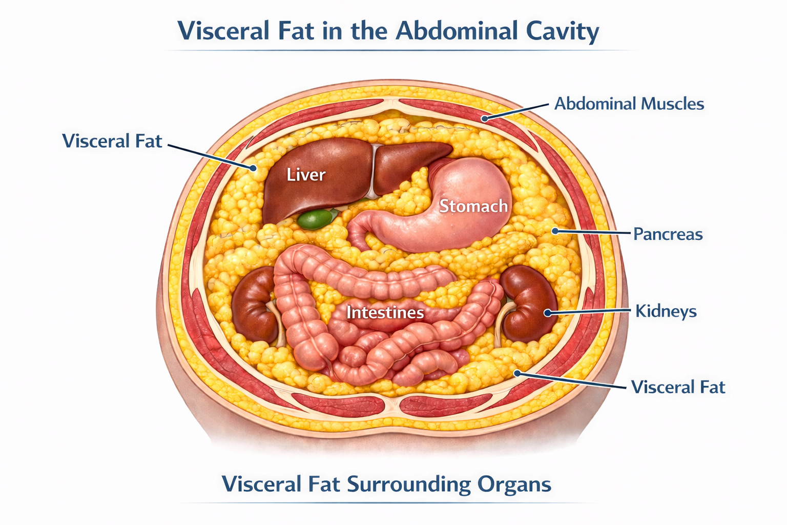
The fat you can pinch just beneath the skin is called subcutaneous fat. It's not metabolically inert, but it's relatively benign compared to the fat that accumulates deep inside the abdominal cavity — surrounding the liver, pancreas, and intestines. That's visceral fat, and it behaves very differently.
Visceral adipose tissue is biologically active. It functions, in many respects, as an endocrine organ — secreting hormones, driving inflammation, and interfering with the metabolic systems that govern energy, appetite, and hormonal balance. The technical term for these secreted compounds is adipokines, and their effects ripple throughout the body.
When visceral fat is elevated, leptin — the hormone that signals satiety — becomes dysregulated, contributing to persistent hunger even in the context of adequate intake. Adiponectin, a hormone that improves insulin sensitivity and reduces inflammation, declines. Pro-inflammatory cytokines like TNF-α and IL-6 rise, creating a state of chronic low-grade systemic inflammation that quietly drives cardiovascular risk, metabolic dysfunction, and hormonal disruption.
Visceral fat is also rich in aromatase — an enzyme that converts androgens like testosterone into estrogens. This is not a minor detail. It means that excess visceral fat doesn't just accumulate as a downstream effect of hormonal imbalance; it actively worsens that imbalance, accelerating androgen breakdown and feeding back on the very hormonal systems that keep fat distribution and metabolism healthy.
Visceral fat doesn't just respond to hormone imbalance — it creates it.
The Hormones Driving Visceral Fat Accumulation
Visceral fat and hormonal dysfunction reinforce each other. Addressing one without the other is rarely sufficient. Here's what the evidence shows for each major hormone axis:
Testosterone
Low testosterone is both a cause and a consequence of visceral fat accumulation. Testosterone promotes lean muscle mass and healthy fat distribution. As levels decline — which happens gradually in both men and women with age — fat preferentially shifts toward the abdomen. That visceral fat then accelerates testosterone breakdown through aromatization, producing more estrogen and suppressing the signaling pathway between the brain and the gonads. The result is a self-reinforcing cycle that worsens over time without intervention.
Estradiol
In women, estradiol plays an underappreciated role in metabolic health. It promotes favorable fat distribution, supports insulin sensitivity, and reduces systemic inflammation. The decline of estradiol at perimenopause and menopause is one of the primary drivers of the visceral fat accumulation many women notice in their 40s and 50s — even without meaningful changes in diet or activity level. Restoring physiologic estradiol is a legitimate metabolic intervention, not merely a quality-of-life measure.
DHEA
DHEA is the most abundant circulating steroid hormone in the body, and it declines significantly with age. Low DHEA correlates with increased visceral adiposity, reduced insulin sensitivity, and an elevated inflammatory state. It receives less attention than testosterone or estradiol in mainstream medicine, but it's a meaningful part of a comprehensive hormonal assessment.
Thyroid — Specifically Free T3
Thyroid hormone drives thermogenesis, fat oxidation, and insulin sensitivity. The clinically relevant form is Free T3 — the metabolically active fraction. Many standard workups stop at TSH, missing patients whose Free T3 is suboptimal even when TSH appears normal. When Free T3 is low, the metabolic engine slows: fat oxidation decreases, insulin resistance worsens, and visceral fat accumulates even in patients doing everything else right.
Cortisol
Visceral adipocytes have a high concentration of glucocorticoid receptors, making them exquisitely responsive to cortisol — the body's primary stress hormone. Chronic psychological or physiological stress translates directly into central fat accumulation, elevated blood glucose, and worsening insulin resistance. Cortisol dysregulation isn't optional to address in any serious approach to metabolic health.
Insulin Resistance
Insulin resistance and visceral fat are so tightly intertwined that separating cause from consequence is often impossible. Visceral fat drives insulin resistance through adipokine dysregulation, chronic inflammation, and excess free fatty acid release into the portal circulation. Insulin resistance, in turn, creates a hormonal environment that favors further visceral fat accumulation. Addressing one without the other rarely produces durable results.
What We Measure — and Why It Matters
Identifying visceral fat burden and its downstream metabolic effects requires looking beyond a scale or a BMI table. BMI, in particular, tells you almost nothing about where fat is distributed or how metabolically active it is. Two people with identical BMIs can carry dramatically different metabolic risk.
At Precision Hormone Consulting, a comprehensive assessment includes:
Waist circumference and waist-to-hip ratio are simple but meaningful starting points — far more predictive of metabolic risk than weight or BMI alone. In-office, we use InBody bioelectrical impedance analysis to go further, generating a validated estimate of visceral fat, lean mass, and body composition that gives us a quantitative baseline and a way to track changes over time. The clinical gold standards for visceral fat measurement — DEXA and MRI — offer greater precision but are expensive and largely inaccessible outside of research settings. For the purposes of clinical monitoring, our combination of anthropometric measures and InBody analysis provides a practical, actionable picture of visceral fat burden without requiring a radiology referral.
Fasting insulin and HOMA-IR — the most direct available measures of insulin resistance. A fasting glucose in the normal range can mask significantly elevated insulin levels, which is where the metabolic damage is already occurring.
Triglyceride/HDL ratio — an accessible and underutilized surrogate for insulin resistance and small, dense LDL particle burden. A standard lipid panel showing normal total cholesterol can coexist with substantial cardiovascular risk in a patient with visceral adiposity and insulin resistance.
hs-CRP — high-sensitivity C-reactive protein, used as a marker of systemic low-grade inflammation driven by visceral fat.
Adiponectin — an inverse marker of visceral fat and insulin resistance. Low levels indicate significant metabolic risk even before glucose dysregulation becomes overt on a standard chemistry panel.
SHBG (Sex Hormone Binding Globulin) — low SHBG is a reliable early signal of hepatic insulin resistance, often appearing before other markers become abnormal. It is particularly useful in women as an early warning sign of metabolic dysfunction.
LH/FSH ratio — in reproductive-age women, normal physiology produces an FSH level approximately twice that of LH. When insulin resistance is present, this ratio begins to narrow — sometimes approaching 1:1 — even before other metabolic markers are overtly abnormal. This is an early, underutilized signal of insulin's effect on the hormonal axis, and it does not require a PCOS diagnosis to be clinically meaningful.
These markers, taken together, provide a far more complete picture of metabolic health than any single value.
The PHC Approach: Treating the Root Cause
A patient came to us in their mid-forties — lean by BMI standards, active, eating well. Their complaint was persistent abdominal fullness and fatigue that had been gradually worsening for two years. Standard labs from their primary care physician had come back normal. Our panel told a different story: suboptimal Free T3, low SHBG, an elevated fasting insulin consistent with early insulin resistance, and a testosterone level that was technically within the reference range but well below what we'd expect for their age and activity level. Within four months of a targeted protocol, their body composition had shifted meaningfully and their energy had returned.
That kind of presentation is common. The tools to identify and address it are available — they just aren't part of routine care.
Hormone Optimization
Restoring physiologic hormone levels is one of the most effective metabolic interventions available. Testosterone optimization in both men and women improves lean muscle mass, reduces visceral fat, and enhances insulin signaling. Estradiol replacement — particularly relevant around perimenopause and menopause — shifts fat distribution favorably and supports metabolic function. DHEA optimization reduces inflammation and supports body composition. Ensuring Free T3 is in an optimal range, not merely a "not flagged" range, restores the metabolic rate that drives fat oxidation.
Peptide Therapy
Growth hormone-releasing peptides are a valuable adjunct for patients with significant visceral fat burden. Tesamorelin has demonstrated specific efficacy in visceral fat reduction in clinical trials. CJC-1295/Ipamorelin combinations support broader growth hormone axis optimization, improving body composition, sleep quality, and recovery.
GLP-1 Medications
For patients with significant metabolic burden or insulin resistance, GLP-1 receptor agonists represent one of the most effective pharmacologic tools currently available. Their mechanisms go well beyond appetite suppression — they improve insulin signaling, reduce hepatic fat, lower systemic inflammation, and produce meaningful, sustained reductions in visceral adiposity.
It's worth noting that GLP-1 is itself a peptide hormone produced naturally in the gut. In patients with metabolic dysfunction, endogenous GLP-1 production is often impaired — meaning these medications are, in a meaningful sense, optimizing a hormone that the body is no longer producing adequately. That framing fits squarely within a hormone optimization model rather than a weight loss drug model.
Lifestyle Integration
No clinical protocol works in isolation. Resistance training, protein-adequate nutrition, quality sleep, and deliberate stress management all independently reduce visceral adiposity and improve insulin sensitivity. Our role is to help patients optimize the full picture — not simply prescribe and monitor. Clinical intervention amplifies the results of good lifestyle fundamentals; it doesn't replace them.
If You've Been Doing the Right Things and Still Not Getting Results
The frustration of doing everything by the book and still carrying stubborn abdominal fat is real — and it usually means something in the hormonal or metabolic picture hasn't been identified yet.
Visceral fat is not a character flaw. It is a metabolic and hormonal problem, and it responds to metabolic and hormonal solutions. The evidence is clear: optimizing testosterone, estradiol, DHEA, thyroid, and metabolic markers produces real, measurable improvements in body composition and long-term health outcomes.
At Precision Hormone Consulting, we specialize in exactly this kind of comprehensive, root-cause evaluation. If you're ready to understand what's actually driving your metabolic health — and address it systematically — we'd be glad to have that conversation.
Schedule a free consultation at precisionhormoneconsulting.com, or call the clinic to book an appointment. Virtual and in-person options are both available.
[DISCLAIMER] This content is for educational purposes only and does not constitute medical advice. Hormone optimization, peptide therapy, and GLP-1 medications involve prescription therapies that require individualized evaluation, monitoring, and ongoing clinical oversight. Some therapies discussed may be used off-label. Results vary. Consult a qualified physician before beginning any new treatment protocol.
Your Mitochondria May Be the Missing Piece: An Introduction to Methylene Blue
You're optimizing hormones, sleep, and nutrition — but something still feels off. The answer may be at the cellular level. A Texas physician explains what methylene blue actually does, who it's for, and why a 150-year-old compound is generating serious clinical interest again.
When You’re Doing Everything Right and Still Feel Wrong
You’re exercising consistently, sleeping reasonably well, eating a clean diet, and managing your hormones with the help of a knowledgeable provider. By most measures, you’re doing what you’re supposed to do. But there’s still something off — a persistent drag on your energy, brain fog that won’t fully clear, or a ceiling on your performance that no amount of lifestyle optimization seems to lift.
In many of these cases, the answer isn’t more optimization at the lifestyle level. It’s happening deeper — inside your cells, at the level of the mitochondria responsible for producing the energy that powers everything else. And there’s a compound with a 150-year track record in medicine that is increasingly being used to address exactly that problem.
It’s called methylene blue. It turns your urine blue. And it’s worth understanding.
What Is Methylene Blue, and Why Does It Have Such a Long History?
Methylene blue is a synthetic compound first developed in 1876 as an industrial dye — the same chemistry behind blue denim. Within a decade it was being used medically: first as a biological stain, then as the first synthetic antimalarial, then for urinary tract infections and psychiatric conditions. It has been formally registered with the FDA for over a century and is on the World Health Organization’s list of essential medications. Its two current FDA-approved indications are treatment of methemoglobinemia and use as a surgical visualization dye. Everything discussed in this post beyond those uses is off-label — applied based on emerging research, and best guided by a physician familiar with the literature. There are nearly 30,000 published studies on PubMed. It is not fringe. It is, however, underused in clinical practice — which is why most patients have never heard of it.
The Mechanism: What Methylene Blue Actually Does in the Body
Mitochondria generate ATP — the currency your body uses for virtually every biological process — through a series of protein complexes called the electron transport chain (ETC). When that chain is impaired, the downstream effects are broad: fatigue, cognitive fog, slow recovery, impaired cellular repair.
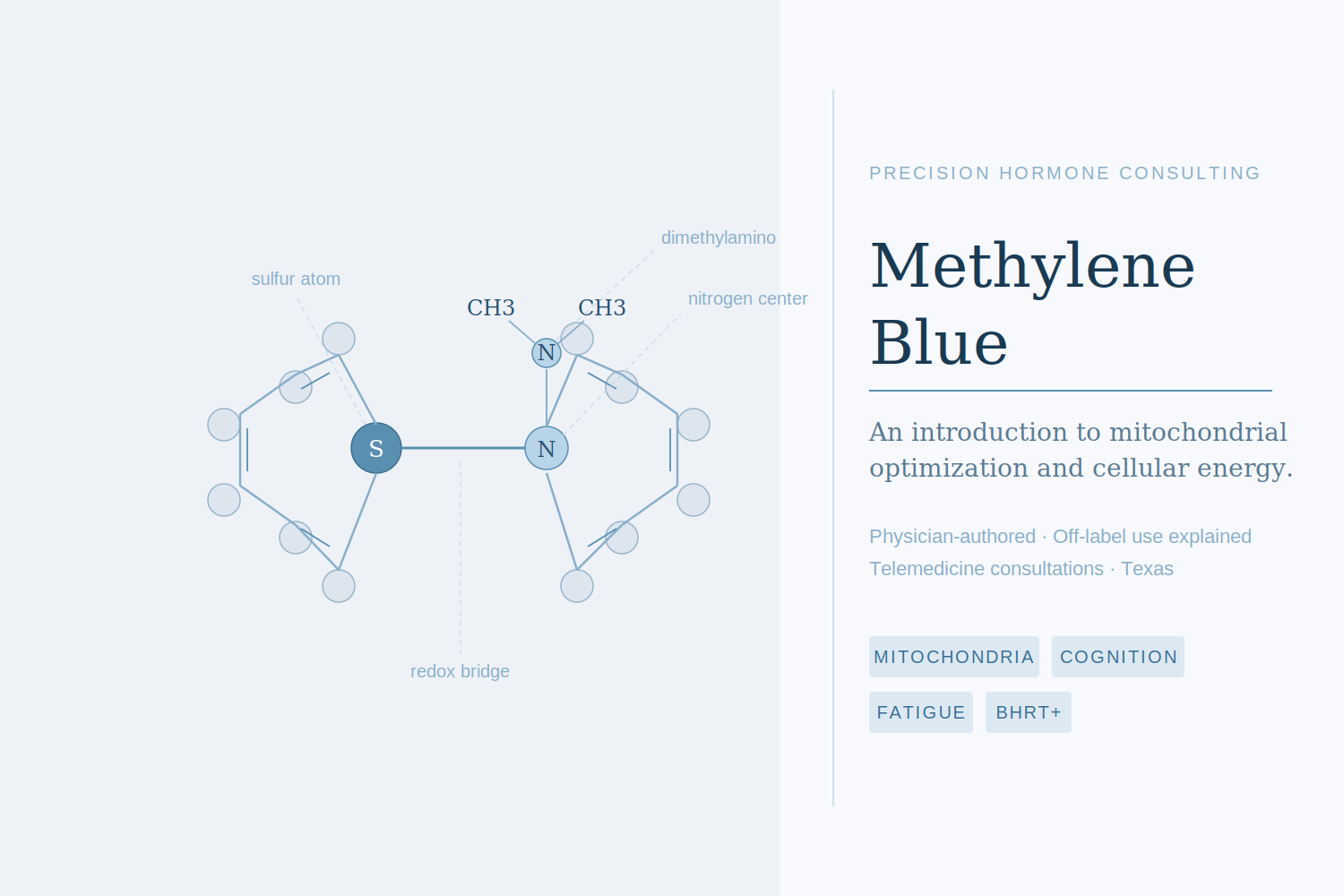
Methylene blue is a redox-active compound that can both accept and donate electrons. In dysfunctional mitochondria, it acts as an alternative electron carrier — stepping in to shuttle electrons past the problem areas and restore ATP production. Think of it as a detour around a blocked highway.
It also scavenges excess reactive oxygen species (ROS), the unstable molecules generated by dysfunctional mitochondria that damage cellular structures. That action is targeted directly at the mitochondria — meaningfully different from a general antioxidant supplement. And at a longer-term level, methylene blue has been shown to upregulate pathways involved in creating new mitochondria, activating PGC-1α and potentially sirtuins, the same longevity-associated proteins linked to exercise and caloric restriction.
The core of methylene blue’s value isn’t one specific condition. It’s the mitochondria — and mitochondrial health is foundational to energy, cognition, hormonal function, and recovery.
Where the Clinical Interest Is Concentrated
Given its mechanism, it’s not surprising that methylene blue is being studied and used across a range of conditions that share mitochondrial dysfunction as an underlying feature. A few areas where the evidence is most developed:
Brain Health and Cognitive Function
The brain is one of the most mitochondria-dense organs in the body, and it is acutely sensitive to disruptions in ATP production. Methylene blue crosses the blood-brain barrier readily, concentrating in neuronal mitochondria. Research has demonstrated increases in brain-derived neurotrophic factor (BDNF), improvements in memory consolidation — particularly fear extinction memory — and anti-apoptotic effects that protect neurons from stress-related damage. In psychiatry, it has a century-long history of use in mood disorders and has shown particular promise in bipolar disorder, including antidepressant and anxiolytic effects without triggering manic episodes.
Fatigue and Post-Viral Syndromes
Chronic fatigue — including the subset of patients dealing with long COVID sequelae — involves measurable mitochondrial impairment and a shift toward inefficient cellular energy metabolism. Methylene blue has been studied for its ability to counteract this metabolic shift, modulate the inflammatory signaling that drives persistent symptoms, and support recovery of normal energy production. Dosing in this context is typically low and titrated gradually.
Dysautonomia and POTS
In conditions characterized by dysregulated vascular tone — including POTS and certain presentations of dysautonomia — methylene blue’s ability to modulate nitric oxide signaling and restore vascular tone has drawn clinical interest. By inhibiting excess nitric oxide production, it can help blood vessels constrict appropriately, improving circulation and reducing orthostatic symptoms.
A Patient’s Experience
A patient in their mid-forties with Hashimoto’s thyroiditis, chronic fatigue, recurrent headaches, and exercise intolerance had thyroid labs being managed but continued to feel limited. After G6PD screening came back normal, methylene blue was added at 50mg daily.
At six months, fatigue and headaches had improved meaningfully and the patient was exercising regularly — something they had not been able to tolerate before. Thyroid antibodies, which had been elevated, normalized. By one year, the patient weaned off methylene blue; the improvements held. This kind of response isn’t guaranteed, but it reflects the pattern that draws providers toward methylene blue in complex, multi-system cases where standard approaches have plateaued.
Who Is a Reasonable Candidate — and Who Should Avoid It
Methylene blue is appropriate for some patients and contraindicated for others. A thoughtful workup before prescribing includes reviewing current medications and screening for specific conditions.
Absolute contraindications include pregnancy (methylene blue is teratogenic), breastfeeding, and G6PD deficiency. G6PD is a genetic enzyme deficiency that impairs the red blood cell’s ability to handle oxidative stress; in affected individuals, methylene blue can trigger hemolytic anemia. G6PD deficiency is more prevalent in people of African, Mediterranean, and South Asian descent, and enzyme levels should be checked before initiating therapy in any patient.
Methylene blue is a monoamine oxidase inhibitor (MAOI), which means it interacts with serotonergic medications — SSRIs, SNRIs, tricyclic antidepressants, and other MAOIs. The risk of serotonin syndrome in the literature is almost entirely associated with high-dose IV administration in surgical settings, not oral use at the doses used in functional medicine. That said, any patient on serotonergic medications warrants a careful, individualized discussion before starting.
Patients who are typically good candidates: those with unexplained fatigue that persists despite optimized hormones and lifestyle, cognitive complaints including brain fog or memory concerns, post-viral syndromes, dysautonomia, or complex multi-system presentations with a suspected mitochondrial component.
Dosing, Purity, and What to Expect
Dosing follows a low-and-slow approach. A typical starting point is 8–16 mg per day, titrated upward based on response — most protocols in this setting land between 15 and 50 mg daily. Purity matters significantly: industrial and chemical grades of methylene blue can contain heavy metals. We prescribe USP pharmaceutical-grade only, sourced through compounding pharmacies with third-party batch testing.
Our formulation pairs methylene blue with ascorbic acid, which improves absorption and moderates the blue urine discoloration — a predictable, harmless side effect worth mentioning upfront. Other dose-dependent side effects include mild nausea or GI discomfort, which typically resolve with dose adjustment. Monitoring begins with a baseline G6PD level and medication review, with follow-up labs tailored to the individual.
Is Methylene Blue Worth Exploring for You?
If you’ve done the foundational work — hormones, sleep, nutrition, exercise — and there’s still a ceiling you can’t break through, the answer may be at the cellular level. Methylene blue is one of the more interesting tools in functional medicine precisely because it addresses a root mechanism rather than a symptom. But like any prescription compound, it deserves a proper evaluation and individualized dosing, not a one-size approach.
At Precision Hormone Consulting, we take the time to understand the full picture before adding anything to a patient’s protocol. If you’re curious about whether methylene blue might be appropriate for you, we’re happy to have that conversation. Free consultations are available virtually — you can book online — or by calling the clinic to schedule in person. No commitment, just a conversation.
[DISCLAIMER]
This content is for educational purposes only and does not constitute medical advice. The use of methylene blue for indications beyond FDA-approved applications is off-label and should only be undertaken under the supervision of a licensed physician who can evaluate your individual health history, current medications, and appropriateness for therapy. Do not start, stop, or change any medication or supplement based on information in this post.